People

Visitor of the Week: Archie Reyes

cshl-visitor-archie-reyes

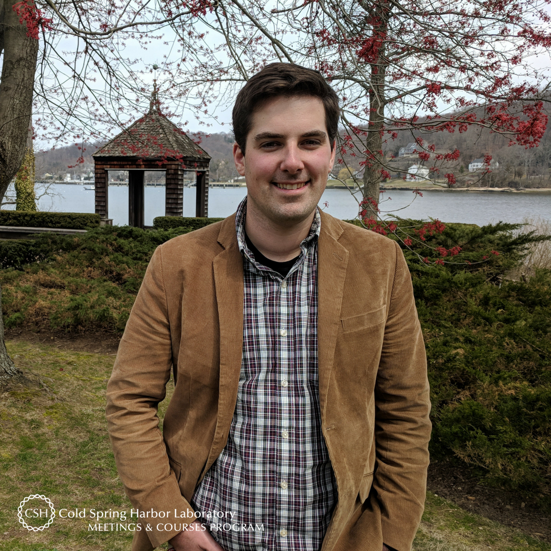
Meet Archie Reyes of the University of Buffalo. The Filipino national is a postdoctoral fellow in John Richard’s lab in the Department of Chemistry. Archie recently made his inaugural trip to CSHL to attend Expression, Purification & Analysis of Proteins & Protein Complexes, and he already has his eyes set on two other courses. 

What are your research interests? What are you working on?
I am interested in enzymes and their modes of action; and studying the general mechanisms on how they provide rate acceleration on several biological processes.

How did you decide to make this the focus of your research? 
While taking my master’s degree, I became really interested in organic reaction mechanisms and protein structure and function and, upon earning my degree, I was sure that I wanted to work in both fields. I realized that the best way to that was to study enzymes – nature’s biological catalysts.

How did your scientific journey begin? 
I have really loved science since I was a kid. It has always been my favorite subject, and I enjoy learning through experiments. My high school chemistry teacher inspired me to start a career in science, specifically in biochemistry.  

Was there something specific about the Expression, Purification & Analysis of Protein & Protein Complexes course that drew you to apply?
There are three reasons why I applied to the course: 1) Learn about other protein expression systems, such as insect and mammalian cells; 2) Be more engaged in proteomics; and 3) Expand my skills in protein purification focusing on protein-affinity tags, as this would help me address our research lab’s need for scaled-up enzyme preparations.

What and/or how will you apply what you've learned from the course to your work? 
I received a lot of help and suggestions from my course instructors and course mates, especially on choosing a more suitable expression system for my enzyme of interest and the appropriate protein-affinity tag to improve the purity of my enzyme preparation.  

What is your key takeaway from the course?
There is so much to learn on purifying and analyzing proteins, and how they interact with other biomolecules. It is important to be familiar with protein biochemisty as it is not just a field of study but also a tool for advancing all life sciences.

How many CSHL courses have you attended?
This is my first time attending a CSHL course, and I am interested in attending either this fall’s X-Ray Methods in Structural Biology or next year’s Cryoelectron Microscopy course.

If someone curious in this course asked you for feedback or advice on it, what would you tell him/her?
CSHL is the best place to learn and obtain new scientific skills in the field of life sciences. If given the chance, I would attend a course from this institution once or twice a year.

What do you like most about your time at CSHL?
I love taking pictures and was able to squeeze in some time to photograph the historically picturesque CSHL! I would love to come back during the fall season to see the institution’s majestic foliage colors.

Archie received a scholarship from the National Cancer Institute (NCI) to cover a portion of his course tuition. On behalf of Archie, thank you to NCI for supporting and enabling our young scientists to attend a CSHL course where they expand their skills, knowledge, and network.

Thank you to Archie for being this week's featured visitor. To meet other featured scientists - and discover the wide range of science that takes part in a CSHL meeting or course - go here.

Visitor of the Week: Jennifer Landino

cshl-visitor-jennifer-landino

Meet Jennifer Landino of the University of Michigan. Jennifer is a postdoctoral fellow and member of Ann Miller's lab in the Department of Cellular, Molecular and Developmental Biology. She was on campus to train at the Cell & Developmental Biology of Xenopus, her very first CSHL course. 

What are your research interests? What are you working on?
I am interested in investigating how cells divide in epithelial tissues, where they are attached to neighboring cells by cell-cell junctions. I specifically want to understand how the neighboring cells contribute to successful cell division while maintaining tissue integrity. 

How did you decide to make this the focus of your research? 
During my PhD research I studied how the cytoskeleton drives cell division in the context of a single cell. For my postdoc, I wanted to expand my understanding of cell division and investigate how groups of cells work together in a tissue to ensure successful division.  

How did your scientific journey begin? 
I became interested in cell division as an undergraduate working on cytokinesis in yeast. This is a fundamental biological process that underlies disease progression, in particular cancer, and has motivated me to continue studying cell division throughout my graduate and postdoctoral training. 

Was there something specific about the Cell & Developmental Biology of Xenopus course that drew you to apply?
My lab uses Xenopus laevis as a model system to study epithelial cell division in a developing animal. I have no experience working with live animals, or developing embryos. This course was a chance to learn about the diversity of experimental techniques that are available in this model system. 

What and/or how will you apply what you've learned from the course to your work? 
I feel more confident in my experimental skills. I also found that my conversations with the instructors and guest lecturers helped me generate ideas that I can use for my own research when I return to the lab. 

What is your key takeaway from the course?
This course was an excellent opportunity to learn and practice a broad range of techniques. I enjoyed learning about the current research in the field - ranging from developmental biology to cell biology. 

If someone curious in attending this course asked you for feedback or advice on it, what would you tell him/her?
This course is a great opportunity to expand your understanding of the Xenopus field. My primary focus is on cell biology, and I have very little experience with developmental biology. I was pleasantly surprised how much I enjoyed learning about classic embryology experiments that were done in Xenopus

What do you like most about your time at CSHL?
 I most enjoyed feeling like part of the Xenopus community. I now feel like I have people I can turn to, outside of my own lab, who will be able to help me grow professionally and scientifically. 

Jennifer received a scholarship from the National Institute of Child Health and Human Development (NICHD) to cover a portion of her course tuition. On behalf of Jennifer, thank you to the NICHD for supporting and enabling our young scientists to attend a CSHL course where they expand their skills, knowledge, and network.

Thank you to Jennifer for being this week's featured visitor. To meet other featured scientists - and discover the wide range of science that takes part in a CSHL meeting or course - go here.

Visitor of the Week: Douglas Reilly

VOTW (2).png

Meet Douglas Reilly of Worcester Polytechnic Institute. The graduate student is in Jagan Srinivasan's lab in the Department of Biology and Biotechnology. He is on campus for the Neuronal Circuits  -- his first CSHL meeting -- where he presented a poster entitled “A neuropeptide controls the sex-specific valence of a mating cue pheromone”.

What are your research interests? What are you working on?
I am interested in how the nervous system encodes instinctive behaviors, and how these responses evolve and diverge in closely related species. I am currently working on understanding how male C. elegans nematodes sense and response to a mating pheromone.

How did you decide to make this the focus of your research?
Going into graduate school, I knew that I wanted to study how the nervous system functions. The Srinivasan Lab’s approach to understanding functional connectomes was something that really grabbed my interest and never really let go.

How did your scientific journey begin? 
In college, my Principles of Neuroscience class finally showed me where to focus my interest in biology. Being able to do an independent study in Prof. Michele Lemons’ lab Assumption College exposed me to what research was like in the neuroscience field. 

Was there something specific about the Neuronal Circuits meeting that drew you to attend?
It’s always great when you have the opportunity to present your work to related fields, and get input from people with related, but very different mindsets. Though there weren’t a ton of worm people at this meeting, getting to talk to fly and mouse neuroscientists increased my confidence in what I was doing, and gave me great ideas regarding the different directions I can take my project.

What is your key takeaway from the meeting?
That the more we learn about the brain, the more we learn how much we don’t know. There were a bunch of fascinating talks which delved into the functioning of well-studied regions of the brain, only to discover previously unknown cells, circuits, and roles within those regions. But even as we realize that we don’t know as much as we think we do, the attendees weren’t disheartened, but instead were only more motivated to continue their work.

Did you pick up or learn something new from the meeting that you plan to apply to your work?  
Discussions with other worm lab researchers here pointed me in the direction of technologies which may help me avoid trouble with completing my project and getting the data published.

If someone curious in attending a future iteration of this meeting asked you for feedback or advice on it, what would you tell him/her?
This meeting has a lot of people doing really cool research on neuronal circuits in a variety of models. But what’s great is that everyone is able to communicate with everyone else, and the ideas exchanged will strengthen your own research.

What do you like most about your time at CSHL?
The ease of talking to people from different backgrounds was great. Unlike topic meetings, this CSHL meeting was not as clique-ish. Everyone was more than happy to talk to anyone else, with fun conversations happening everywhere and between people who had just met.

Thank you to Doug for being this week's featured visitor. To meet other featured scientists - and discover the wide range of science that takes part in a CSHL meeting or course - go here.

Visitor of the Week: Alisha Geldert

cshl-visitor-alisha-geldert

Meet Alisha Geldert, a PhD student in the UC Berkeley-UCSF Graduate Program in Bioengineering.  Alisha is a member of Amy Herr's lab in UC Berkeley and is on campus taking part in her first course at CSHL: Quantitative Imaging: From Acquisition to Analysis.

What are your research interests? What are you working on?
I am interested in developing tools to help biologists and/or clinicians make quantitative measurements on biological samples. Currently, I am working on increasing the precision of a single-cell Western blotting assay developed in my lab, which enables high-throughput measurements of protein expression from single cells.

How did you decide to make this the focus of your research? 
I am excited by the ways new biological measurement tools can expand research capabilities and improve diagnostics. I am intrigued by what we may be able to learn from single-cell measurements, because measuring with this increased level of resolution can reveal rare subpopulations of cells or unique cell states which could have important implications in disease.

How did your scientific journey begin? 
In general, I had always enjoyed my math and science classes, but my interest in pursuing a career in bioengineering really developed as I began to see how math and science could actually be used to improve healthcare. My interest developed through anything from watching a How It’s Made episode on contact lens design, to creating a simple ECG device in a laboratory course, to seeing the limitations of medical care that my friends and family have encountered.

Was there something specific about the Quantitative Imaging: From Acquisition to Analysis course that drew you to apply?
Because my lab focuses on developing new biological measurement tools, quantitative fluorescence imaging and analysis are crucial components of my research. I applied to this course because I wanted to better understand not just the microscope itself, but also the many other factors (illumination sources, detectors, image analysis techniques, etc.) that affect the accuracy and precision of quantitative fluorescence measurements.

What is your key takeaway from the course?
Every microscopy technique has tradeoffs, so you must have a clear understanding of the scientific question you are trying to answer with microscopy and consider the tradeoffs of different imaging and analysis techniques before settling on an experimental plan.

What and/or how will you apply what you've learned from the course to your work? 
I really appreciate that the course instructors have framed the material in a practical manner – they teach about different types of microscopy in the context of how to design experiments with the proper controls and validation so that measurements are more likely to be meaningful. Since my lab owns a few microscopes of our own (rather than performing all imaging in a core facility), lab members share responsibilities for maintaining and troubleshooting issues with our microscopes, and this course has better prepared me for this task.

How many CSHL courses have you attended?
This is my first course at CSHL and I would love to come back! For the past few years or so, my lab has taught a portion of the CSHL course on Single Cell Analysis so perhaps I will get involved in that course in the future.

If someone curious in attending a future iteration of this course asked you for feedback or advice on it, what would you tell him/her?
I would definitely recommend it, especially for early-stage graduate students, because you will develop a solid fundamental understanding of quantitative microscopy that can improve your experimental design and troubleshooting skills throughout the course of graduate school.

What do you like most about your time at CSHL?
I have really enjoyed getting to know the other course participants, teaching assistants, and instructors. It is a very diverse and fun group!

Alisha received a scholarship from the Helmsley Charitable Trust to cover a portion of her course tuition. On behalf of Alisha, thank you to the Helmsley Charitable Trust for supporting and enabling our young scientists to attend a CSHL course where they expand their skills, knowledge, and network.

Also thank you to Alisha for being this week's featured visitor. To meet other featured scientists - and discover the wide range of science that takes part in a CSHL meeting or course - go here.

A Word From: Florin Albeanu, Michael Orger, Lucy Palmer & Philbert Tsai

L to R: Ken Zaret, Fiona Watt, Marius Wernig; Photo by Constance Brukin

Last summer, the twenty-seventh  annual course on Imaging Structure & Function in the Nervous System was held at Cold Spring Harbor Laboratory. We chatted with the 2017 co-instructors Florin Albeanu, Michael Orger, Lucy Palmer, and Philbert Tsai to gain an inside look into the long-running, intensive course  First, we asked about a day-in-the-life of an Imaging trainee, and learned they literally build microscopes from scratch during the three-week course: 

Phil: The typical daily schedule changes from week to week but overall, we try to avoid an information overload. For example, in the first week, we don’t like to bombard the trainees with a lot of information all at once so the schedule is kind of lecture, practical, lecture, practical. The practicals are lab bench exercises that allow the students to tinker with stuff and effectively learn how to build a microscope from scratch. 
We begin at 9 AM with three hours of lectures on the fundamental of optics and methods of microscopy to ensure everyone has a good foundation of the basics. After lunch, the students spend two to three hours working on optical bench rails and building small imaging systems. The systems become more complicated with each practical, from simple lens imaging to a full wide-field microscope to a laser scanning microscope (which is a bonus for some students because it can be turned into a confocal microscope). Later in the afternoon, we have another lecture that is oftentimes followed by questions that came up during the practical. There are questions the students will only ask if they’ve actually been confronted with the fact that something doesn’t work the way they thought it would; if we jumped from one lecture to the next without a practical in between, those questions would not come up and the depth of conversations would not be the same. By the end of the first week, the students move into the main lab and start working with million-dollar microscopes, at which point they have a better idea of what’s going on behind the lens. 
Michael: As the students transition from the optical bench exercises to the microscope, the first step is to conduct experiments designed to better understand the fundamental principles of what they’ll do in their own work. They measure the noise characteristics and resolution of the microscopes so they understand the possibilities and limitations in their experiments in a very general way. When they’re using the microscopes in the course, they’re really learning how to understand the measurements they’re making and what they mean.
Florin: During the second and third week of the course, there are a lot of applications of the basic ideas taught in the first week. And in many cases, the trainees realize that exactly the same concepts are packaged in different microscopes. Fancy up-and-coming microscopes, in fact, use the same two or four lenses again and again. 
Lucy: In the third week, the students also work on their own course projects, which they can pursue solo or as part of a group.
Michael: The final assignment of the course, right before the lobster banquet, is for the students to present the results of their projects or anything else they want to share regarding what they learned during the course. 
Phil: Outside of the course projects, we always have them work in at least pairs because we feel the social aspect is important. When the students do the optical bench exercises, they work in groups of 3 or 4 which indirectly becomes a team building exercise. A majority of students never thought that they could build a microscope. So there’s this team effort to overcome the hurdle of “We’re supposed to build a microscope from scratch in three hours? No way!” And they all end up doing it. 
Florin: They also realize that the microscopes they build, in many cases, can do as good a job as the pricey microscopes on the market. That knowledge goes a long way. If funding is a limitation, they have the experience to build their own microscope and understand exactly how everything works in it. 
Phil: The microscopes the students build use $40 lenses and $50-100 scanners, so they do have certain limitations. They’ll never be as good as a well-engineered, million-dollar microscope, but a homemade version can get you 95% of the way there. The critical thing is realizing what that last 5% is and whether it’s worth hundreds of thousands of dollars. When you’re doing cutting edge research, it’s important to understand where the critical components are so you know where to spend your money and where not to waste it.
Florin: Getting the students to play with the lenses themselves actually makes this much more obvious. More than that, as you look at the microscope, you realize the precision of the microscope is the precision of your hands, of your alignment, and that’s empowering. 
Michael: The students also have the opportunity to try new things and test out ideas. We have a combination of commercially built equipment--a lab of ten of the nicest microscopes at their disposal--and equipment just for developing and trying new things. Very often, actually, the course projects aim to make a particular method a little bit better. 
Phil: Microscopy is one of those topics that falls between traditional departments. On the physics side, the basics of how a microscope is constructed are taught but most physics departments don’t teach their undergraduates about laser scanning microscopes or confocal microscopes. Biologists, on the other hand, use these microscopes all the time but are unfamiliar with the working principles behind the microscopes. Over the years, alumni have told us they don’t know where else they could have learned all this, that the course is the only place they could find all this information.
Florin: Also, a physics book on optics can be a little dry. So if you are a biologist trying to understand imaging, learning through a book can be a long path. Being part of a course like this exposes you to all kinds of tools, principles, and techniques in optical imaging, then you actually use them to build a device.
Michael: You can read a book about optics but until you get your hands on things, it’s really hard to fully grasp the concepts.

Invited speakers play a major role in CSHL courses, including the Imaging course. Next, the instructors discussed the caliber of the 2017 speakers and their contributions to the course and its trainees. 

Michael: We schedule the invited speakers on a bit of a gradient: speakers at the beginning of the course cover basic methodologies and those toward the end talk about more specific applications. Our speakers are really, really great. A number of them have been coming to the course for over a decade, possibly even two.
Phil: And they evolve. Their talks are always on their most recent research as well as what they’ve done in the past. We invite them back because they enjoy this crowd and its interactive-ness. These are conference-level speakers who give colloquia to departments or universities in front of hundreds of people. The course trainees get to be in the room with them, have lunch with them, and ask them whatever questions they have. That’s a real opportunity.
Michael: It’s a completely different experience than if the person came to your university and gave a talk. In the course, the students can really ask the speakers whatever they want. And almost all of the speakers like to stay at the course for extended periods to spend time with the students.
Phil: Many of our speakers are the ones who started this field.  
Florin: Sometimes new projects arise from these interactions; course alumni have joined the labs of invited speakers they met in the course.
Phil: Let me phrase it this way: it’s the chance for an interaction that sometimes changes the course of a career. It’s the types of conversations that are had in a crowd of 12 people, in an atmosphere that encourages questions, ideas, and curiosity. There have been times when a student might ask, “I’m interested in X. Could X be used for Y?” And when a person who created X and uses it every day answers with, “Actually, X would be ideal for Y,” it’s very encouraging. The student follows a new research direction because of that one exchange. 

We then asked how the course has changed over years:

Lucy: Phil is probably the best person to answer since he’s been with the course for 15 years.
Phil: I’m the best and worst person to answer this because I have a terrible memory! Fundamentally, I think the course hasn’t changed that much over the last 15 years. This biggest evolution has been fine-tuning the iterative back-and-forth of listening to lectures, followed by tinkering in the lab practicals, followed by more lectures and Q&A sessions. I think we’re in a pretty good place with that now. 
Lucy: The course content also evolves as new approaches come out. We really try to stay on top of the field with different preps and techniques, so the students are always exposed to cutting-edge material. 
Phil: More and more students are coming to the course with the word ’microscopy’ already in their background. Ten to fifteen years ago, students applied to the course because imaging was becoming big and was gaining momentum in the field of neuroscience – but not everybody was yet doing it. Today, imaging is starting to dominate. You’ll be hard-pressed to find a core facility or university without a two-photon microscope. As a result, the students now have some prior two-photon imaging or confocal imaging experience, and they want to learn what’s going on inside the microscope when they adjust settings. So the things we teach in the first week haven’t changed much. And despite students having done microscopy at their home lab before arriving at the course, they still gain quite a bit from the material taught the first week. 
Michael: I think there’s been an increased focus on in vivo preparation. When I was a student of this course in 1999, there was a lot of cell culture and various other preps. But imaging activity on the cortex of a mouse was not something we did.
Phil: It was more limited to in vivo slices and cell cultures.
Lucy: Synthetics.
Florin: Funnily enough, things that were talked about in the course 10 to 15 years ago as dream projects are actually being done now in the class by students. And we still talk about “the new dream experiment” that will probably be implemented in the next decade by people involved in the course now. 

As one of our most competitive courses, we made sure to inquire how applicants might increase their chances on becoming one of the 14 to 16 scientists each summer who are accepted into the course. 

Michael: We look for applicants who are new to imaging and understand some of the problems they’re facing, but don’t yet fully understand what they’re doing. We want to train people who will be able to return to their labs and quickly take advantage of what they learned in the course. A lot of information is taught during the three weeks so it’s important that students can attend the course, understand why they’re learning what they’re learning, and go back to their labs to directly apply their newfound knowledge. This way, the information is reinforced as soon as possible. 
Florin: We also look for applicants who are about to switch fields or start a new project. 
Michael: We try to accept a mixture of students in terms of scientific background. We have had some students from physics backgrounds who have never seen a biological specimen, but have a firm understanding of how some elements of microscope systems work. And vice versa. It’s great to have a mixture like that because the students learn not just from the instructors, speakers, and TAs, but from each other. They can ask each other questions and share their knowledge. 
Applicants’ personal statements should really express how the course is going to benefit their experiments or, even further, their labs. Of course, we want students with great scientific backgrounds, but the personal statement is less about selling yourself as a scientist and more about how this course is going to help you to do science better. For example, if you’re in a lab that is just starting to set up imaging but doesn’t have an electrophysiologist, that student’s participation in the course will have an impact even beyond their own research projects. 
Another important thing to note is that every year we reject a bunch of great students. If you really want to take this course and think this is what you need, don’t give up. We consider persistence a big positive. So if an applicant states “I applied last year and didn’t get in and now I’m applying again,” it catches our attention. Besides wanting a group of motivated students who are fun to work with and enthusiastic about the experiments, we want people who really want to take the course.

Our chat closed with the co-instructors answering, “What’s your favorite moment from the course?” 

Florin: It was my first encounter with the course. In 2006, I came in as a TA and was supposed to build a device in a short amount of time. Clearly, I was terrified dealing with the parts, but the instructors at that time said, “don’t worry, it’s going to work out just fine.” And indeed it did. Here, you are surrounded by extremely enthusiastic people whose encouragement makes a lot of things happen in a very short amount of time. 
Phil: My favorite moment happens every year and it changes in flavor: it’s that moment when the students, who have been working on a rig for hours, struggling with noise and all sorts of other stuff, finally obtain a fluorescent image. They put together this scanning microscope themselves from a table of random parts and spent an hour aligning it, and now they see a high-quality image. They literally scream and jump for joy. They’re taking pictures of their microscopes and the image with their phones, posting it on Facebook, texting it to their loved ones and colleagues. It’s the excitement of realizing that they’re capable of something they didn’t know they were capable of just a few hours ago. 
Michael: A lot of great moments happen throughout the course but one thing that is a pleasure to see takes place some years after a student’s been in the course. I see them post on Facebook about their new publication, and actually see the imaging they came to the course to learn. It’s always very satisfying to be reminded that the course has an impact on the students beyond just the three weeks. 
Lucy: I have quite a lot of favorite moments from when I was a student in the course, and now as an instructor. As a student, it was building my own two-photon which was amazing: I was elated when I took the image. And now as an instructor, my favorite part is seeing all the students get to that same pinnacle and realizing they can do it. I enjoy seeing the motivation and excitement in students when they’re able to address a question they’ve always wanted to address. 

The Imaging Structure & Function in the Nervous System course this summer will take place between July 24th and August 13th. Applications are being accepted until this Sunday, April 15th here. For an introduction into the course from a trainee, be sure to read our Q&A with 2017 Alumna Janani Sundararajan

For more conversations with other course instructors, check out the rest of our A Word From series.