Quantitative Imaging Course

Visitor of the Week: Temitope "Temi" Adebambo

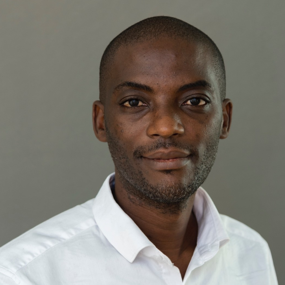
Meet Temitope “Temi” Adebambo of Emory University. The Nigerian citizen is a postdoctoral fellow and member of Dorothy Lerit’s lab. He is participating in the Quantitative Imaging: From Acquisition to Analysis (QICM) – his first course at CSHL and since the pandemic. Temi shared that “there is [a] healing side to having a program in person” and we could not agree more.

Tell us about your research.
My research is focused on how toxins affect development using Drosophila as a model organism. I am also interested in the roles played by centrosome in regulating cell cycle and how this affect the neurobiology of flies.

How did you decide to focus on this area/project?
Trained as a toxicologist, my Ph.D. demonstrated the global effect of low molecular weight aromatics in flies using genome-wide techniques and linking this to cell cycle defects in mitotically active imaginal wing disc.

What and/or who is the inspiration behind your scientific journey?
Prof. Adebayo Otitoloju, my undergrad/Masters/Ph.D. degrees mentor has been an excellent inspiration for my scientific journey.

What impact do you hope to make through your work?
Teaching and research is my thing and I want to do so much research, acquire relevant training in order to provide meaningful mentorship for the next generation of researchers.

What do you love most about being a researcher?
No time spent in conducting research is ever wasted, there is always a lesson to be learnt.

What drew you to apply to this course?
To be a better researcher but most importantly to help me better settle into cell biology.

What is your key takeaway from the Course; and how do you plan to apply it to your work?
Important microscopic techniques that were learned by accident or by necessity in a haste are no longer so. I have a better knowledge of the behind-the-scene operations of these techniques and how to tailor them to my research interests. From Brightfield to TIRF microscopes and Deep machine learning, I now have the right combination of tools to advance my research.

What feedback or advice would you share with someone considering to participate in this course?
If you think you know microscopy, you don’t.

What’s the most memorable thing that happened during the Course?
I have never seen a live cell imaging system before--that is forever memorable.

What do you like most about your time at CSHL?
I don’t have to worry about food; I don’t have to cook and there is absolutely no need to rely on any transport system as everything is in one location. Whoever came up with the idea to have a discussion every morning before lectures and the little break in between lectures is a genius. Those are always very helpful.

Thank you to Temi for being this week's featured visitor. To meet other featured researchers - and discover the wide range of science that takes part in a CSHL meeting or course - go here.

Image provided by Temitope Adebambo
Visitor of the Week: Chad Hobson

VOTW (4).png

Meet Chad Hobson of the University of North Carolina at Chapel Hill (UNC-CH). A second-year graduate student in the physics program at UNC-CH and member of Richard Superfine’s lab, Chad is at CSHL training at the Quantitative Imaging: From Acquisition to Analysis course thanks in large part to a fellowship from the Helmsley Charitable Trust and support from the Graduate Student Opportunity Fund at UNC-CH.

What are your research interests? What are you working on?
My main interest is in designing new systems that combine cutting-edge microscopy techniques with force-measurement instruments to investigate the mechanics of single cells. Specifically, I am using a combined light sheet microscope and atomic force microscope to investigate the mechanics of cell nuclei.

How did you decide to make this the focus of your research?
I toured Rich Superfine’s lab during an undergraduate summer program (REU) at Duke University and loved the idea of combining force and imaging techniques and fundamental physics to understand the mechanics of single cells. The field was entirely new to me, but the ability to design and invent as well as use my physics background was inspiring.

How did your scientific journey begin?
During my freshman year of undergraduate school at Lynchburg College, my professor, Dr. John Eric Goff, approached me and asked if I would be interested in conducting sports physics research under his guidance. I started work with him in the summer before my sophomore year and have not looked back. I have changed gears from sports physics to nuclear physics to now biophysics, but my passion for research has remained unchanged.

Was there something specific about the Quantitative Imaging: From Acquisition to Analysis course that drew you to apply?
I really wanted to get hands-on experience with a variety of imaging techniques in order to both broaden my general knowledge of microscopy as well as understand the difficulties and limitations of each technique. These skills will help increase the rigor and success of my graduate research going forward.

What and/or how will you apply what you’ve learned from the course to your work?
Everything I have learned directly applies to our lab. Whether it is general maintenance and care of optics or a more detailed understanding of each component of a microscope, I am excited to share what I have learned with my fellow lab researchers.

What is your key takeaway from the course?
The biggest takeaway so far is that I will never know everything about microscopy, and that is okay. At the course, however, we are building up from the basics so that I can develop a working knowledge that applies to almost all imaging modalities. Moving forward if I need to understand the detailed intricacies of a certain method I have the foundation to do so.

If someone curious in attending this course asked you for feedback or advice on it, what would you tell him/her?
Take it! The instructors are beyond fantastic and helpful, and you will never look at a microscope the same way again.

What do you like most about your time at CSHL?
I have truly enjoyed getting to know the fellow students and instructors at the course. We all come from such different backgrounds and fields, so learning about what brought us all here together has been an enlightening experience. 

Chad received funding support via a fellowship from the Helmsley Charitable Trust and from the Graduate Student Opportunity Fund at UNC-CH. On behalf of Chad, thank you to these organizations for supporting and enabling our young scientists to attend a CSHL course where they expand their skills, knowledge, and network.

Thank you to Chad for being this week's featured visitor. To meet other featured scientists - and discover the wide range of science that takes part in a CSHL meeting or course - go here.

Repeat Visitor: Leila Rieder

Photo provided by Leila Rieder

Photo provided by Leila Rieder

The next scientist to be featured in the 2018 edition of our Repeat Visitor series is Leila Rieder. Leila is a K99 postdoctoral fellow in Erica Larschan’s lab in Brown University with a visiting appointment at Princeton University. And, next April, she will take on her new role as Assistant Professor in the Biology Department of Emory University. Leila made her CSHL Meetings & Courses debut in August 2017 when she trained at the Proteomics course. She returned for this year’s Quantitative Imaging: From Acquisition to Analysis course (QICM) followed by a poster presentation at the Nuclear Organization & Function meeting a few weeks later. We caught up with Leila to chat about her experience at our meetings and courses, and if she has any plans of participating in a near future meeting or course.

Tell us about your research interests and how you decided to make it the focus of your research.

I’m most interested in how genes are coordinately regulated -- turned on and off at the same time. Cells are remarkably good at this and we don’t know how they do it! I first began by focusing on how sex chromosomes are singled out for unique regulation, a process called dosage compensation. In these systems, basically all the genes carried on a single chromosome are coordinately regulated. However, it’s not simply the chromosomal location that leads to this coordinated regulation; there are other signals. While researching the role of a known dosage compensation protein on the male X-chromosome, I discovered the same protein was involved in regulating another group of coordinated genes: the histones. These genes are often clustered together within genomes and are notable and unique for many reasons. Every time a cell divides, it needs a huge output of histone proteins in the right ratios so they are incredibly important genes!

How did your scientific journey begin?

My father is a cell biologist and, to be honest, because it was important to me to forge my own path, I tried very hard to be anything other than a biologist! But because of my father’s research, I spent my childhood summers at the Marine Biological Laboratory and sat through so many meals where the entertainment (the entire conversation!) was dominated by my father and his colleagues debating their newest results. They couldn’t get together without “talking science” and I wanted to have those conversations myself -- and now I do!

Quantitative Imaging: From Acquisition to Analysis Class of 2018

Quantitative Imaging: From Acquisition to Analysis Class of 2018

This year, we hosted 26 meetings and 34 courses - how did you narrow down which ones to attend?

I taught myself most of the microscopy I know, and was intensely aware that I was probably doing 50% of it incorrectly. (After the course, I realized I was closer to 90%.) I knew that if I wanted to perform experiments correctly, I needed formal guidance and a background in theory. I’ll soon be in a position to guide my trainees through their own research, and will make sure to never suggest they do sloppy or ignorant microscopy!

As for the meeting, I decided it was important to understand how my favorite genes and loci were situated in the nucleus. I had been working in the field of coordinated gene regulation but without deeply considering higher-order organization and genomic context.

What is your key takeaway from QICM? Also, what advice would you give to someone interested in this course?

Quantitative microscopy is incredibly powerful when used correctly, but there are many ways to use it to find false positives and red herrings. Most of all, I will never apply a quantitative microscopy technique without first researching all the ways in which it can go wrong. I think the best time to take this course is when you have some basic microscopy experience but haven’t yet significantly applied it to your biological question. Be prepared to hear about the different ways your past experiments were sub-par. It’s not a great feeling, but it’s better to learn sooner rather than later!

For Nuclear Organization & Function, what feedback can you provide for those interested in participating in its 2020 iteration?

Leila during her poster presentation at the 2018 Nuclear Organization & Function meeting.

Leila during her poster presentation at the 2018 Nuclear Organization & Function meeting.

The field of nuclear organization and function is much broader than I imagined! It focused on specific topics that were not as relevant to my work as I had expected but, due to it, I now read more broadly. The meeting itself is a great opportunity to meet people—both those who are everyday names as well as those you might not have heard of but are doing really interesting work. I presented a poster entitled “Dynamic identification of the dosage-compensated Drosophila male X-chromosome during early embryogenesis,” and the experience was intense, as many poster sessions are, but friendly. I liked that the meeting was small so it was neither difficult to find people nor for them to find me.

Since you’ve experienced both meeting and course life at CSHL, did you notice any differences or similarities between the two function types?

Since you basically live and work with your course mates for two straight weeks, you really get to know them. We come from so many backgrounds and different countries, and are using what we learn during the course for wildly different purposes. This diversity really adds to the experience. This is all true of the meeting participants as well, but you don’t get to know them to the same extent since the meetings are only four days long.

Our readers are always eager to learn of ways to fund registration. Can you share how you were able to fund your CSHL meeting and course participation?

Since I am located at Brown and Princeton Universities - neither of which are far from CSHL - transportation to CSHL is easier and less costly for me than for most. For tuition and registration support, I received small grants from my home institution, NCI for QICM, and CSHL generously delayed payment for the course tuition until my K99 grant was available. I was also provided tuition support by NICHD when I took the Proteomics course in 2017.

What did you like most about your experience at CSHL?

Overall, I enjoyed meeting so many interesting people from backgrounds so different than mine. At the meeting, what I liked the most was the chance to sit next to someone new at dinner. As for the course, the instructors, TA’s, and vendors were really fantastic and you can absolutely tell they love teaching the course—they live and breathe it even more than the students do! And they had boundless energy. They never got tired of answering questions and, when the students finally shuffled out at the end of a long evening, they stayed to set up for the next day. They are amazing!

Do you have a future CSHL course or meeting on your radar?

Yes! Now that I am about to begin my own research group, I plan on taking the Workshop on Leadership in Biosciences this coming March. And, someday if I have time, I’d like to take the Programming for Biology course. I’m also planning to attend the Mechanisms of Eukaryotic Transcription meeting in 2019.

Both the Quantitative Imaging: From Acquisition to Analysis and Proteomics courses will return to the Laboratory in 2019; and applications are already being accepted. Apply to QICM by January 31, 2019 here, and to Proteomics by April 1, 2019 here.

Thank you to Leila for sharing with us her experience, and we look forward to having her back at the Laboratory again. To meet other featured scientists - and discover the wide range of science that takes part in a CSHL meeting or course - go here and here.

A Word From: Jennifer Waters & Talley Lambert

c-qicm-18

The annual CSHL course on Quantitative Imaging is one of our most in-demand courses, attracting -- this year alone -- four times as many applications as it has spots. The course debuted in 2011 as a reinvention of a previous long-running course on immunocytochemistry and live-cell imaging. 

Last month, we sat down with two of the four co-instructors, Jennifer Waters and Talley Lambert, both of whom were instrumental in shaping what the course has become today. Because it’s so competitive, our chat began with how they select trainees each year from the pool of applicants: 

Jennifer: I go right to the personal statement: I want to know what their motivation is for taking the course. We’re focused not just on microscopy, but on using microscopes as a tool for making measurements – quantitative microscopy – and we’re looking for applicants who recognize the importance of that in their research. We also look for applicants who we think, after having taken the course, will benefit others. So, for example, an application from a lab that develops fluorescent proteins but doesn’t have a lot of classically-trained microscopists would be a great candidate; having a microscopist in that group would be a great help in validating the proteins. 
Talley: We like to make sure that what they are seeking is a good match for what we're teaching. 
Jennifer: That’s true. Sometimes people are looking for something different. We also want applicants who, based on where they are in their career, will benefit the most from the course. We often get applications from first-year graduate students and, based on our many years of teaching, we know that’s a bit too early. They have to have their hands in research for a while. Second-year graduate students are borderline. We do take them, and we had a couple this year, but I think third year is the sweet spot. By then, they’ve really figured out what their project is, and they’ll take what they learn here and start using it right away.
Talley: I definitely like re-applications too.
Jennifer: Yes! We have gotten applications we would like to accept but didn’t because we thought they weren’t quite ready yet. So we’re happy when we see those people apply to the course again; in fact, the chances of their re-application being selected is higher. 

If you’re among the ~25% accepted to train at this course, this is what your typical day would look like: 

Jennifer: It’s exhausting. We start at 9 o’clock sharp and for an hour and a half, we have coffee and continental breakfast while going over questions and reviewing anything the trainees didn’t quite get during the previous night’s lab. To me, it’s really important to have a good stretch of time each day for open discussions. After that, we launch into the first lecture of the day, which tends to run for another hour and a half, sometimes two hours. Next is lunch and then it’s another lecture, lab, or both in the afternoon. With the exception of maybe one evening talk, we try not to schedule any lectures after dinner. Instead, we end the day in the lab, and that generally goes until 10 o’clock. 
Talley: There’s very little unscheduled time from 9 AM to 10 PM, except one hour for lunch and one hour for dinner.
Jennifer: If a person has never been involved in this sort of course, then it might feel like a lot. But compared to other courses, we don’t go as late into the evenings; I just think people need a good night’s sleep if they’re going to continue to learn while they’re here. Even though the pace is intense, there’s a lot of time where we’re just talking because the labs are very much set up like that. The TAs and instructors all genuinely enjoy teaching, so they’re in the lab, checking if anyone has questions, chitchatting, getting cornered by the trainees to talk about individual projects at home, etc. So there’s a lot of time when we’re just interacting and getting to know each other, to the point where we’re all sad on the last day that it’s over. 
Talley: For anybody we bring in as a TA or invited speaker, I see it as a requirement that they be there for the students, because it’s fun and necessary for them to get access to experts. 
Jennifer: I agree, it’s really important for the trainees to get as much of our time as they want or need. 

The discussion then moved to how the course has and hasn’t changed in the last eight years:

Jennifer: Well, Talley and I haven’t changed – we’re still here! [Laughter]
We have always had an analysis component, which is one of the things that makes our course unique. We are serving what we see as a great need: people who want to use microscopy and do analysis need to learn both of them. That’s a very hard thing to do because each topic could easily fill the full two weeks of the course. 
But the course has changed quite a bit since 2011. For one, a majority of the lectures are now given by people like Talley and me -- people who run core facilities. Our jobs consist of talking to and guiding people who need to use microscopy and use it properly. We do a lot of teaching at home, so we have built up an expertise and understand what the course trainees don't know, and what they need to know. We put a lot of practice into the best ways of presenting concepts so trainees can absorb the ridiculous amount of material we throw at them in a short period of time. We do still have professors and principal investigators, who are absolute experts in their specific applications, come to the course and give seminars where they show really nice, rigorous applications of the type of technology that we teach. 
Something else that has changed is the technology itself, and we make an effort to keep up with it and stay cutting-edge. For example, we didn’t teach light sheet microscopy at all the first couple of years but as it is now coming of age, we’ve doubled the amount of lecture and lab-time spent on it. Each year, we think of what new technology people should know about – even if they're not asking for it. 
Talley: In any given year, I can think of things we've added or removed. We used to put more time into two-photo microscopy but have sort of de-emphasized that a bit. We also used to have a sample preparation component that is completely gone now, and we've emphasized super resolution imaging to varying degrees. The different microscopy techniques just kind of ebb and flow, and it's a continuous consideration to change with them.

Besides being a lead instructor for this course, Jennifer is also a lecturer at two of our other courses, Drosophila Neurobiology and Imaging Structure & Function in the Nervous System. We asked her about differences between the three courses, and if there’s a benefit for a Drosophila or Imaging alum to also train at the Quantitative Imaging course:

Jennifer: Absolutely. Both the Drosophila and Imaging courses are designed for imaging a specific application. The Drosophila course is all about how to prepare flies for imaging and then how to image them. The Imaging course is a little broader in that it’s imaging for neuroscience, but it’s still very much focused on that field. So they  spend a lot of time on multi-photon imaging, which we don’t, because that’s commonly used in neuron imaging. In our course, we don’t play favorites in terms of what organism you want to image, and we cover a wide range of imaging modalities that can be used. In fact, somebody who has taken one of the other two courses actually has a great background for then taking our course. We like people to have a little bit of microscopy experience for our course because there’s a lot of material that takes a lot of time to absorb.

We next asked for their favorite moments from the course over the past eight years:

Talley: When I think of our highlights, I think of the guest lecturers. 
Jennifer: Oh! We had Eric Betzig one year before he won the Nobel Prize. 
Talley: That’s a good highlight.
Jennifer: I like to think it means that we’re really good at picking guest lecturers. [Laughter]
Talley: Correlation and causality. [More laughter]
Jennifer: The year he won the Nobel Prize, it was on my list to invite him again. Not because he’d just won but because he’s great and really fascinating to listen to. He kindly declined of course, but I had to email him anyways. I’m always very proud of the lecture series we put together for the course. Our invited speakers are microscopists who speak at big meetings in front of large auditoriums of people. For them to come here, give a lecture to our 16 students, have dinner and chat with them, it’s such a great experience. 
Talley: This is not a specific moment, but when one of the students asks an invited speaker a great question that you’re pretty confident they just learned at the course, and yet they totally get it…
Jennifer: That pride. 
Talley: Yeah, and sometimes a student gives a presentation on something they did in the course lab that is really striking and very, very good. Just seeing the trainees’ progress. It’s not a specific moment but it’s definitely a category that we always talk about.

Like Jennifer, Talley has been with the course since 2011. However, Talley started out as trainee, then worked his way through the “ranks” to his current role as co-instructor. Here’s what he had to say about his evolution with the course:

Talley: It’s difficult to overstate the impact of taking this course. I mean, it changed my career. I came as a biologist to learn some techniques and left saying, “I think I want to do that instead.” I was just enamored with all of it. 
In the beginning, it was just all learning and I didn’t really feel like I had anything to give. I was absolutely terrified when I gave my first lecture, and it was on-camera! Now I feel like I have a lot to give -- I feel genuinely capable of fielding any topic in this course. The biggest change has been in my level of confidence and the relative degree of what I’m learning versus what I’m giving. It’s been fantastic. I enjoy teaching this course and I love Cold Spring Harbor because this is where things changed for me. 

Jennifer concluded our chat with why she continues to teach this course at CSHL: 

Jennifer: I have taught at other places besides Cold Spring Harbor and I love teaching here. The support that we get is amazing. My home institution keeps asking me to run a course like this there; I said I’d consider it if they give me a team of people like the one Cold Spring Harbor has to support this course!

The Quantitative Imaging: From Acquisition to Analysis course returns to the Laboratory next April and is already accepting applications here. For an inside look into the course, visit its Twitter and Facebook accounts. 

For more conversations with other course instructors, check out the rest of our A Word From series. 

Photo: Constance Brukin

Visitor of the Week: Alisha Geldert

cshl-visitor-alisha-geldert

Meet Alisha Geldert, a PhD student in the UC Berkeley-UCSF Graduate Program in Bioengineering.  Alisha is a member of Amy Herr's lab in UC Berkeley and is on campus taking part in her first course at CSHL: Quantitative Imaging: From Acquisition to Analysis.

What are your research interests? What are you working on?
I am interested in developing tools to help biologists and/or clinicians make quantitative measurements on biological samples. Currently, I am working on increasing the precision of a single-cell Western blotting assay developed in my lab, which enables high-throughput measurements of protein expression from single cells.

How did you decide to make this the focus of your research? 
I am excited by the ways new biological measurement tools can expand research capabilities and improve diagnostics. I am intrigued by what we may be able to learn from single-cell measurements, because measuring with this increased level of resolution can reveal rare subpopulations of cells or unique cell states which could have important implications in disease.

How did your scientific journey begin? 
In general, I had always enjoyed my math and science classes, but my interest in pursuing a career in bioengineering really developed as I began to see how math and science could actually be used to improve healthcare. My interest developed through anything from watching a How It’s Made episode on contact lens design, to creating a simple ECG device in a laboratory course, to seeing the limitations of medical care that my friends and family have encountered.

Was there something specific about the Quantitative Imaging: From Acquisition to Analysis course that drew you to apply?
Because my lab focuses on developing new biological measurement tools, quantitative fluorescence imaging and analysis are crucial components of my research. I applied to this course because I wanted to better understand not just the microscope itself, but also the many other factors (illumination sources, detectors, image analysis techniques, etc.) that affect the accuracy and precision of quantitative fluorescence measurements.

What is your key takeaway from the course?
Every microscopy technique has tradeoffs, so you must have a clear understanding of the scientific question you are trying to answer with microscopy and consider the tradeoffs of different imaging and analysis techniques before settling on an experimental plan.

What and/or how will you apply what you've learned from the course to your work? 
I really appreciate that the course instructors have framed the material in a practical manner – they teach about different types of microscopy in the context of how to design experiments with the proper controls and validation so that measurements are more likely to be meaningful. Since my lab owns a few microscopes of our own (rather than performing all imaging in a core facility), lab members share responsibilities for maintaining and troubleshooting issues with our microscopes, and this course has better prepared me for this task.

How many CSHL courses have you attended?
This is my first course at CSHL and I would love to come back! For the past few years or so, my lab has taught a portion of the CSHL course on Single Cell Analysis so perhaps I will get involved in that course in the future.

If someone curious in attending a future iteration of this course asked you for feedback or advice on it, what would you tell him/her?
I would definitely recommend it, especially for early-stage graduate students, because you will develop a solid fundamental understanding of quantitative microscopy that can improve your experimental design and troubleshooting skills throughout the course of graduate school.

What do you like most about your time at CSHL?
I have really enjoyed getting to know the other course participants, teaching assistants, and instructors. It is a very diverse and fun group!

Alisha received a scholarship from the Helmsley Charitable Trust to cover a portion of her course tuition. On behalf of Alisha, thank you to the Helmsley Charitable Trust for supporting and enabling our young scientists to attend a CSHL course where they expand their skills, knowledge, and network.

Also thank you to Alisha for being this week's featured visitor. To meet other featured scientists - and discover the wide range of science that takes part in a CSHL meeting or course - go here.